ULTRASTRUCTURE OF BACTERIAL CELL
ULTRA-STRUCTURE OF
TYPICAL BACTERIAL CELL:-
Under electron
microscope the structure of bacterial cell is look like a capsule. It has
various components. Some of these are outside the cell membrane;
others are inside of cell membrane.
The outer layer or cell
envelope consists of two components such as cell wall and a cytoplasmic or
plasma membrane. Inside the plasma membrane, there is protoplasm comprising the
cytoplasm, cytoplasmic inclusions such as ribosomes, mesosomes, granules,
vacuoles, and nuclear body.The cell may be enclosed in a viscid layer which is
termed as capsule. Many bacteria have filamentous appendages called fimbriae or
pili (organ of adhesion). Many bacteria also posses flagella which are organs
of locomotion.
Components outside the cell membrane:-
1. Capsule
2. Pilli
3. Flagella
4. Cell wall
Components inside the cell wall:-
1.
Cytoplasmic membrane (Plasma membrane)
2.
Cytoplasm
3.
Ribosomes
4.
Mesosomes
5.
Intracytoplasmic inclusion
6.
Nucleoid & nucleus
7. Spores
1. CAPSULE:-
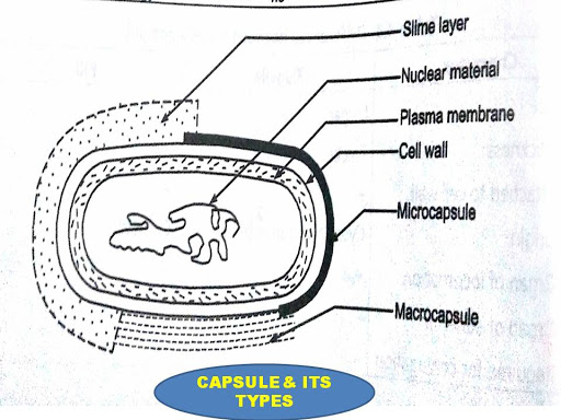
It is the outer layer of the bacterial cell. Many prokaryotic micro organisms synthesize
amorphous organic exopolymers which are deposited outside the cell wall called
capsules or slime layer or glycocalyx or sugar coat.
Capsule refers to the layer tightly attached to
the cell wall. While the
Slime layer
is the loose structure that often diffuses into the growth medium.
Depending
on the chemical nature capsule are thick or thin.
Microcapsule:-
Capsule layer may be thin of a size less than 0.2 µm called microcapsule.
Macrocapsule:-
Thick layer of size more than 0.2 µm to 10 µm called macrocapsule.
Capsules
may be composed of a complex polysaccharide (Klebsiella pneumoniae) polypeptide
(Bacillus anthrocis) hyaluronic acid
(Streptococcus pyogenes). Water (98%) is the main component of bacterial
capsule. Capsule or slime layer has less affinity for battle dyes and is not
visible in Gram staining. Special capsule staining techniques are used by using
copper salts as mordants. Capsules may be easily observed by negative staining
in wet films with lndian ink.
Functions of capsule:
1.
They may provide protection against mechanical
injury, temperature & temporary drying by binding water molecules.
2.
They may promote attachment of bacteria
to surfaces. E.g. Streptococcus mutant that cause dental carries attach on teeth
surface by its capsule.
3.
They may promote the stability of
bacterial suspension by preventing the cells from aggregation and settling.
They inhibit
phagocytosis (antiphagocytic) and contribute to the virulence of pathogenic
bacteria.
PILLI/FIMBRIAE:
-
Many Gram negative bacilli
contain short, thin, hair-like microfibrils called Pilli. The size of the pilli is 0.5 to 2 um in length
and 5 to 7 nm in diameter.
They are more numerous than flagella. Fimbriae are composed of protein known as
pillin and its molecular weight is 18000 daltons. Fimbriae can be seen only under the electron
microscope. They are best developed in freshly isolated strains and in liquid
cultures. They may disappear after sub culturing on solid media.
FUNCTIONS OF PILI:-
1.
Pilli are non motile but adhesive structure.
They enable the bacteria to stick firmly to other bacteria & to a surface hence
pili is also called an organ of adhesion.
Pili is used for the
transfer of genetic material from the donor to the recipient cell (bacterial
conjugation).
FLAGELLA:-
Flagella
are long, slender, thin hair like structure. The size of the flagella is about 0.01
µm to 0.02 µm in diameter & 3 to 20 µm in length. Flagella are made up of a
protein (flagellin) similar to keratin or myosin & they are responsible for
the motility of bacteria hence it is called as organs of locomotion.
Flagella
are found in both Gram positive and Gram negative bacteria. A large no of bacteria
such as spirilla, vibrios, most of bacilli & few coccal forms, are motile
by means of flagella.
Flagella
can be seen by an ordinary light microscope by special staining techniques in
which their thickness is increased by mordanting.
Flagellum
has three basic parts (1)
Filament, (2) Hook and (3) Basal body.
Filament is the thin, cylindrical, long outermost region with a
Constant diameter. The protein in the filament is made up of monomers called
‘flagellin' with molecular weight ranging between 20,000 to 40,000 The filament
is attached to a slightly wider hook, consisting of a different protein
. The basal body is composed of a small central rod inserted into a
series or rings.
Gram-negative bacteria
contain four rings as L-ring, P-ring, S -ring and M ring. L-ring is embedded in Lipo-polysaccharide layer of outer membrane membrane, P-ring in Peptidoglycan layer, S-ring in just above cytoplasmic membrane (Semi-position of
membrane) and M-ring within cytoplasmic Membrane. Gram-positive bacteria have
only S and M rings in basal body.
The
number and arrangement of flagella are characteristics of each bacteria.
Flagella may be seen on bacterial body in the following manner.
- MONOTRICHOUS:-
These bacteria have single polar flagellum.
E.g. vibrio cholera, pseudomonas aeruginosa, spirillum.
- LOPHOTRICHOUS:- Bacteria have two or more flagella only at one end of the cell. E.g. pseudomonas fluorescens.
- AMPHITRICHOUS:- Bacteria have single polar flagella at both poles. E.g. Alcaligenes fecales, Aquaspirillum serpenes.
- PERITRICHOUS:- Several flagella present all over the surface of bacteria. E.g. E.coli, Salmonella typhi.
CELL WALL:-
Cell wall is a rigid structure which gives definite
shape to the cell and protect from osmotic lysis.
They are situated between the capsule and plasma
membrane.
SIZE:- It
is about 10 20 nm in thickness and constitutes 20-30% of the dry Weight of the cell. The wall can
protect a cell from toxic substances and is the site of action of several
antibiotics.
Peptidoglycan-the most
important molecule in the cell walls of bacteria. Peptidoglycan or murein is an enormous
polymer composed of many identical subunits. The polymer contains two sugar
derivatives, N-acetylglucosamine and Nacetylmuramic acid (the lactyl ether of
Nacetylglucosamine), and several different amino acids, three of
which—D-glutamic acid, D-alanine, and meso-diaminopimelic acid
Gram + cell wall:- The gram-positive cell wall consists of a single 20 to 80 nm thick.
They are homogeneous peptidoglycan or murein layer lying outside the plasma
membrane. Homogeneous
cell wall of gram-positive bacteria is composed primarily of peptidoglycan,
which often contains a peptide interbridge . The teichoic acids are connected
to either the peptidoglycan by a covalent bond with Nacetylmuramic acid or to
plasma membrane lipids are called lipoteichoic acids.
Gram – cell wall:- The gram-negative cell wall is quite complex. It has a 2 to 7 nm
peptidoglycan layer surrounded by a 7 to 8 nm thick outer membrane. The outer
membrane lies outside the thin peptidoglycan layer • The most abundant membrane
protein is Braun’s lipoprotein, covalently joined to the underlying
peptidoglycan and embedded in the outer membrane by its hydrophobic end. • Constituents
of the outer membrane are its lipopolysaccharides • outer membrane is more
permeable than the plasma membrane due to the presence of special porin
proteins
Functions of cell wall:
- Cell wall is involved in growth and
cell division of bacteria.
- It gives shape to the cell.
- It gives protection to the internal
structure and acts as a supporting layer.
- It provides attachment to complement.
- It shows resistance to the harmful effects of environment.
CYTOPLASMIC MEMBRANE:-
Also called as plasma membrane, is the
most dynamic structure of a bacterial cell. The
cytoplasmic membrane is a thin (5 to 10 nm) layer lining the inner surface of
the cell wall and separating it from the cytoplasm. It is composed Of phospholipids
(20 to 30%) and proteins (60 to 70%).
The phospholipids form a bilayer surrounding the
cytoplasm and regulate the flow of substance in and out of the cell in which
most of the proteins are tenaciously held and are called integral proteins.
Other proteins are loosely attached are called peripheral proteins. The phospholipids
molecules are arranged in two parallel rows. called a phospholipids bilayer.
Each phospholipid molecule contains a polar head composed of a phosphate group
and glycerol. The non-polar tails are in the interior of the bilayer and the
polar heads are on the two surfaces of the phospholipids bilayer.
Functions
of cytoplasmic (plasma) membrane:-
- Its main function
is a selective permeability barrier that regulates the
passage of substances into and out of the cell.
- It provides mechanical strength to the bacterial cell.
- It helps in DNA replication,
segregation with septum formation & cell division.
- It contains the enzyme,
permease, which plays an important role in the passage of selective
nutrients & ions through membranes.
- It contains the enzymes involved in the biosynthesis of membrane lipids and synthesis of murein (cell wall peptidoglycan) & other macromolecules of the bacterial cell
CYTOPLASM:-
The
bacterial cytoplasm is a Gel-like matrix composed of mostly water (4/5 th ),
enzymes, nutrients, wastes, and gases
The cytoplasm of bacteria differs from that of higher eukaryotic microorganisms
as it not contain endoplasmic reticulum, Golgi apparatus, mitochondria and lysosomes.
It contains ribosome, chromosomes, plasmids, proteins as well as
the components necessary for bacterial metabolism. It carries out very important functions for the cell - growth, metabolism,
and replication.
The main constituents of cytoplasm
is Proteins
including enzymes Vitamins, Ions, Nucleic acids and their precursors – Amino
acids and their precursors – Sugars, carbohydrates and their derivatives –
Fatty acids and their derivatives.
RIBOSOMES:-
The most notable structures in the bacterial cytoplasm
are the ribosomes. They are involved in protein synthesis &
translate the genetic code from the molecular language of nucleic acid to that
of amino acids. Bacterial ribosome’s
are similar to those of eukaryotes, but are smaller and have a slightly
different composition and molecular structure. Bacterial ribosome’s are never
bound to other organelles as they sometimes are bound to the endoplasmic
reticulum in eukaryotes, but are free-standing structures distributed
throughout the cytoplasm. Their number
varies with the rate of protein synthesis {15000/cell). The greater the rate of
protein synthesis the greater the number of ribosome’s.
The bacterial ribosomes are referred to as 70S ribosomes.
(S-Svedberg unit, the unit of sedimentation).These ribosome’s when placed in a
low concentration of magnesium, dissociate into two components as 5OS and 30S
particles.
Each
50S particle contains one molecule of 23S-RNA, one molecule of 5S-RNA and 32
different proteins. The 30S subunit contains one molecule of 16S r-RNA and 21 different
proteins. These ribosome’s, during active protein synthesis are associated with
the m-RNA and such associations are called polysomes.
MESOSOMES:-
Mesosomes are also called chondroids and
are visualized only under an electron microscope. Mesosomes
are the invaginated structures formed by the localized infoldings of the plasma
membrane. The invaginated structures comprise of vesicles, tubules of lamellar
whorls. In some bacteria particularly in gram-positive bacteria
depending upon the growth conditions the membrane appears to be infolded at
more than one point such infoldings are called as mesosomes. Generally
mesosomes are found in association with nuclear area or near the site of cell
division. They are absent in eukaryotes.
There
are two types of mesosomes:-
Central mesosomes:-
Are present deep into the cytoplasm and
located near the middle of the cell. This mesosome is attached to bacteria
chromosome and is involved in DNA segregation and in the formation of cross
walls during cell division.
Peripheral mesosomes:-
Are not restricted to a central location
and are not associated with nuclear material.
mesosomes are supposed to take part in respiration but they
are not analogous to mitochondria because they lack outer membrane. In
the vesicle of mesosomes the respiratory enzymes and the components of electron
transport such as ATPase, dehydrogenase, cytochrome are either absent or
present in low amount.
Mesosomes might play a role in reproduction also. During
binary fission a cross wall is formed resulting in formation of two cells.
Mesosomes begin the formation of septum and attach bacterial DNA to the cell
membrane.
In
addition, the infoldings of mesosomes increase the surface area of plasma
membrane that in turn increases the absorption of nutrients.
INTRACYTOPLASMIC iNCLUSIONS:- It is also
called as inclusion bodies. Bacteria can produce within their cytoplasm a variety
of small bodies which is called as inclusion bodies. Some are called granules
and other are called vesicles. They are mainly used for storage of energy &
reduce osmotic pressure by tying up molecules in particulate forms like
polysaccharides granules, glycogen granules, metachromatic granules, lipid
granules etc.
Inclusions are considered to be nonliving components
of the cell that do not possess metabolic activity and are not bounded by
membranes. The most common inclusions are glycogen, lipid droplets, crystals,
and pigments.
Granules:-
metachromatic or
Babes~Ernst granules are highly refractive, basophilic
bodies consisting of polymetaphosphate.
They appear reddish when stained with polychrome methylene blue or
toluidine blue. Albert's or Neisser's special staining techniques are used for
the study of metachromatic granules.
Lipid
granules consist mainly of polymerised beta hydroxybutyric
acid. They can be seen in unstained preparations and by staining with sudan
black and by modified ziehl-Neelsen stain. Polysaccharide
granules can be stained with iodine. They are made-up of either glycogen
(red brown) or starch (blue).
Vesicles:-
Some aquatic photosynthetic bacteria and cyano
bacteria have rigid gas-filled vacuoles and it helps in floating at a certain
level - allowing them to move up or down into water layers with different light
intensities and nutrient levels.
NUCLEOID
AND NUCLEUS:-
The nucleoid is a region
of cytoplasm where the chromosomal DNA (genetic material) is located. Bacterial
nucleus does not possess nuclear membrane, nucleolus and
deoxyribonucleoprotein.
Most bacteria have a single, circular chromosome
that is responsible for replication, although a few species do have two or more
smaller circular auxiliary DNA strands, called plasmids, are also found in the
cytoplasm.
Bacterial
nucleus can be demonstrated by acid or ribonuclease hydrolysis. They may be
seen by a light microscope after staining (Feulgen stain) or by electron
microscopy. They appear as oval or elongated
bodies generally one per cell. The genome consists of a single molecule of
double stranded DNA arranged in a circle. It may open under certain conditions
to form a long chain about 1000 um in length.
SPORES:
-
Many
bacterial species produce spores inside the cell (endospores) as well as
outside the cell (exospores). eg. Bacillus anthracis, Bacillus subtilis,
Clostridium tetani etc.
Endospores
are thick-walled, highly refractile bodies that are produced one per cell. Each
bacterial spore on germination forms a single vegetative cell. Therefore
sporulation in bacteria is a method of preservation and not reproduction.
Spores are extremely resistant to dessication, staining, disinfecting
chemicals, radiation and heat.They help bacteria to survive for long periods
under unfavourable environments.
All
endospores contain large amount of dipicolinic acid (DPA) with 10 to 15 percent
of the spores being dry weight
An
endospore returns to its vegetative state by a process called germination,
which has three distinct stages.
Activation:
The
activation process requires agents like heat, low pH, abrasion etc. These
agents damage the coat of the spore and help in germination by growing in a
nutritionally rich environment.
Initiation:
Binding
of the effector substance (L-alanine. adenosine, glucose etc.) to the. spore
coat, activates an autolysin which destroys peptidoglycan of the cortex.
releases calcium dipicolonic acid and absorbs water.
Outgrowth: Spore coat breaks and
forms a single germ cell. It starts growing into a new vegetative cell and
active synthesis takes place producing an outgrowth.
CLICK BELOW TOPIC TO READ


0 Comments
Please do not enter any spam link in the comment box.